
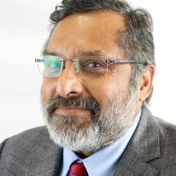
Mr Kartik Hariharan
Orthopaedic Foot and Ankle Surgeon
- Orthopaedic Surgery
- Self-pay/Insured
How does it feel?

Mr Kartik Hariharan
Orthopaedic Foot and Ankle Surgeon
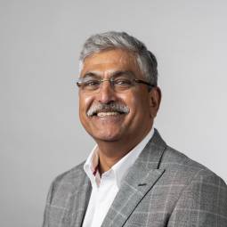
Mr Sujit Kadambande
Orthopaedic Surgeon
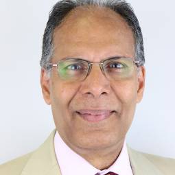
Mr Yogesh Nathdwarawala
Orthopaedic Surgeon

Our transparent pricing and bespoke packages allow you to pay for the treatments and services you need, when you need them.

Many of our dedicated consultants have partnered with insurance companies to give you peace of mind with your health.
For more information call one of our friendly patient advisors or book online using the button below.